
The Common Thread--How Evolution Ensured Its Continuation Down Through the Ages
Episode 3: In which the author digresses from the main story line for a drabble about mtDNA (Everything you ever wanted to know about mtDNA (but were afraid to ask!).

Previous Episode: The story follows Mila returning to camp with two newly caught rabbits, after which the family works together to process the animals, cook rabbit stew in a carefully managed log cooker, and use every part of the catch. It also depicts everyday family life—Grandpa Ara teaching the children to carry and refill water using small bladders, while younger brothers Josen and Balay handle their waste elimination routines and then wash up to rejoin the group.
100-Word Detour:
Survival of the Fittest (Taking on A Whole New Evolutionary Adaptive Meaning)
Cast
Zeke and Xavier Spermatatoa:
Brother sperm taking part in this month’s “race” arrive at the starting gate:
Whoa, Bro’, Ya ready fer dis?
‘sI’ll ever be, I s’pose. You?
Yep, bin trainin’ onna sched’al Hey, you seen da course?
Naw, I reckon dis is the end fer me. Too much comp’tition. Might’s well git it over with. Mebbe come back as a girl next time—cushy life jus’ layin ‘round in d’egg’s cytoplasm.
Awww, don’t be a wuss. I’m takin a chance, Baby. Gonna get me inside dat ovum ‘n’ be da first guy to pass on ‘is mitachondria.
You kiddin,’ bro’? Y’ain’t heard ‘bout wha’ happens to male mitachondria? You might’s well be a praying mantis!
Not really! It’s a myth that the female eats the male after mating; It only happens 30% of the time. AND, there’s very good evolutionary rationale for it, for both male and female, believe it or not! (See below)
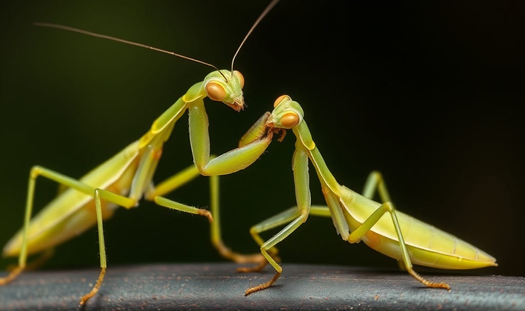
Read on to Learn What Happens to Male Mitachondria. Yep, Even Those Who Get Inside!
Author’s Note: (7.15.26)
I had to learn a few more details before I could keep writing mtMRCA’s story. Here is the convo I had with duck.ai where I got some facts.
Long Conversation with Duck.ai in which I learn about mtDNA
Does the mother pass on any DNA other than mitochondrial DNA?
Yes. A mother passes on far more than mitochondrial DNA (mtDNA).
Mitochondrial DNA: mtDNA is typically inherited almost exclusively from the mother (via the egg), because the embryo’s mitochondria mostly come from the mother’s cytoplasm.
Nuclear DNA (the majority of DNA): the mother passes half of the nuclear chromosomes to her child (about 50%), while the father provides the other half. This nuclear DNA contains most genes and is what determines most inherited traits.
Occasional exceptions: very rarely, mitochondria from the father can occur (reported in some cases), but for most practical purposes mtDNA is treated as maternal.
Me: What particular genes do the mitachondria DNA control?
Mitochondrial DNA (mtDNA) controls a small, specific set of functions inside mitochondria—mainly making parts of the cell’s energy machinery.
In humans, mtDNA genes mainly encode:
13 protein subunits used by the oxidative phosphorylation (OXPHOS) system (the electron transport chain and ATP synthase components).
22 tRNAs (transfer RNAs) used to build those mitochondrial proteins.
2 rRNAs (ribosomal RNAs) used to produce those mitochondrial proteins.
So, mtDNA most directly affects how efficiently mitochondria generate ATP (cell energy)—which then can influence tissues with high energy demand (like muscle and brain).
Me: Do we know how they influence tissues with high enegy needs?
Yes—mechanistically, we have a pretty clear picture of how mitochondrial DNA (via its effects on mitochondria) can influence high–energy tissues.
Mitochondria are the main site of oxidative phosphorylation, where they make ATP. mtDNA encodes key parts of that ATP-making machinery, so changes in mtDNA can shift:
ATP availability: Less efficient electron transport can mean lower ATP production, so tissues that constantly demand ATP (brain, heart, skeletal muscle) can’t meet their energy needs.
Reactive oxygen species (ROS): If the electron transport chain runs less smoothly, electrons can “leak” and form more ROS, which can damage cells and further impair energy production.
Cell stress and signaling: Energy shortfalls and oxidative stress can trigger pathways that alter cell function, promote dysfunction, or—if severe—lead to cell loss.
Heteroplasmy effects (mix of mtDNA variants): Individuals have a mixture of normal and altered mtDNA in their cells. Different tissues may reach a high enough fraction of altered mtDNA to cross a “threshold,” which helps explain why certain tissues are more affected.
Clinically, many mtDNA-related disorders show this tissue pattern (especially neurologic symptoms, muscle weakness/exercise intolerance, and cardiac involvement), consistent with the idea that those tissues are most sensitive to disruptions in mitochondrial ATP production.
Me: Do we have any idea why the mitachondrial DNA ende up only in the ovum?
Yes—there’s a well-supported biological explanation for why mitochondrial DNA (mtDNA) is usually inherited almost exclusively from the mother.
In fertilization, the egg provides almost all the cytoplasm (and therefore almost all the mitochondria) that end up in the embryo. By contrast, the sperm contributes very little cytoplasm, and its mitochondria are packaged in a part of the sperm (the midpiece) that generally doesn’t persist after fertilization.
After the sperm enters the egg, the remaining question is what happens to the sperm’s mitochondria:
In most organisms (including humans), sperm mitochondria are actively removed and/or degraded soon after fertilization.
The egg cell also triggers processes that eliminate those paternal mitochondria, while the egg’s own mitochondria remain.
So the “maternal-only” pattern comes from a combination of:
maternal cytoplasm/mitochondria are what the embryo mainly receives, and
paternal mitochondria are rapidly eliminated.
Me: Hmmmm, How come the ovum came about the process of eliminating the paternal mitachondria? Are we related to the Praying Mantis?
The egg eliminates paternal mitochondria because it has to keep a consistent mitochondrial genome and because, in most species, the fertilizing sperm is mainly delivering DNA, not mitochondria.

How the egg eliminates paternal mitochondria (big picture)
After fertilization, paternal mitochondria are usually marked for removal and then broken down by the egg’s cell-cleanup machinery. The main ideas are:
Selective degradation signals: Components in the egg help identify the paternal mitochondria as “not to be kept,” often via tagging them with proteins that signal destruction.
Autophagy / cellular recycling: Once tagged, the egg can send those mitochondria into autophagy (a lysosome-based “eat and recycle” pathway), so the paternal mitochondria are dismantled and their parts reused.
Egg-dominant persistence: Even without perfect “targeting,” sperm mitochondria generally don’t survive well in the egg’s intracellular environment, and many organisms actively suppress them.
Are humans related to the praying mantis?
No—this is not something mantises “invented” or anything like that. The underlying reason (egg cytoplasm and cell-cleanup pathways controlling what mitochondria persist) is a general reproductive-cell biology theme across many animals, including humans. It evolved separately in different lineages; it’s not a special mantis-only relationship.
Me: That was a joke, ya know?
Got it 🙂—all good.
Even so, your mantis joke fits the biology vibe: in most animals, paternal mitochondria don’t “stick around,” because the egg’s internal cleanup and control systems mostly prevent them from surviving—so mitochondria end up being transmitted from the mother rather than the father.
Me: But why, I wonder? what would the original “trigger” for that mechanism in the original species in which it developed? Something to do with the Y chromosome?
It’s not “triggered” by the Y chromosome (mtDNA elimination happens in many species regardless of Y/X sex systems), and there wasn’t a single ancient “design moment” where an organism decided to remove paternal mitochondria.
A good evolutionary explanation is:
1) There’s a practical advantage to having one mitochondrial lineage.
Mitochondria have their own genome but they must work with mitochondrial proteins made mostly in the nuclear genome. If you allow mitochondria from both parents to coexist, you can create “mismatched” combinations (different mtDNA variants) that may be less compatible with the host cell’s nuclear setup. Keeping transmission mostly maternal reduces that kind of problem.
2) Paternal mitochondria can be treated as “unwanted cargo” because the egg doesn’t need them.
After fertilization, the embryo is already supplied with plenty of mitochondria via the egg’s cytoplasm. Sperm mitochondria are a leftover from the fertilization process, and in most animals they’re either physically limited in quantity and/or get targeted for destruction.
3) Mechanisms that reduce paternal mtDNA can spread over generations if they improve fitness.
Even if early organisms didn’t eliminate paternal mitochondria perfectly, random variation existed in how well paternal mitochondria survived. Any tendency to reduce paternal mtDNA that caused fewer incompatibility issues (or less damage, less competition among mtDNA types, etc) would be favored by natural selection.
So the “original trigger” isn’t a specific Y-related signal—it’s more like selection for eggs that consistently end up with their own mitochondria (or avoid paternal mtDNA persistence) because mixed mitochondrial populations can cause trouble.
Mitochondrial–nuclear compatibility (plain language)
Mitochondria make most of their proteins using two genetic sources:
mtDNA encodes a subset of mitochondrial proteins (mostly the core energy/ATP machinery parts plus RNA pieces to translate them).
nuclear DNA encodes the rest of the mitochondrial proteins (and a lot of the “maintenance” proteins that help run and assemble the energy machinery).
So inside a cell, mtDNA-encoded parts have to work with nuclear-encoded parts that the cell inherited from its parents.
Why mixed mtDNA can be a problem
In most normal reproduction, an offspring gets:
nuclear DNA 50/50 from mom and dad
mtDNA mostly from mom
That means the mitochondrial genome and the nuclear “toolkit” are usually in a co-adapted arrangement.
If an embryo were to inherit substantial mtDNA from both parents, you could end up with:
two different mtDNA types competing inside the same cell (heteroplasmy from mixed sources), and/or
mtDNA variants that are less well matched to the nuclear background that ended up in that embryo.
The practical outcome can be:
less efficient ATP production (energy deficits),
more reactive oxygen species (stress/damage),
or generally poorer mitochondrial performance—especially in energy-hungry tissues.
How this connects to selection
Because mitochondrial performance affects survival and reproduction, embryos/tissues where paternal mtDNA doesn’t persist (or is removed) would, on average, have a fitness advantage if mixed mtDNA combinations were more likely to be harmful.
That creates evolutionary pressure for mechanisms that:
reduce survival of paternal mitochondria, and/or
ensure the embryo ends up dominated by a single mtDNA lineage (usually maternal).
If you’d like, I can also explain what “heteroplasmy thresholds” means and why certain tissues (like muscle/brain) show symptoms more readily.
sure
Heteroplasmy thresholds (what that means)
Heteroplasmy is when a cell (or tissue) has a mixture of mitochondria/mtDNA variants—e.g., the maternal type plus some paternal type (or maternal variants that differ).
Across development, the fraction of the “different” mtDNA can shift a lot because:
mitochondria replicate inside cells,
some mitochondria get diluted or lost during cell division,
different cells end up starting with different mtDNA mixtures.
A threshold means: mitochondrial dysfunction tends to show up only when the harmful (or less efficient) mtDNA fraction gets high enough. Below that, the remaining healthy mtDNA can supply enough energy.
Why tissues show different effects
Different tissues have different energy demands and different “buffers”:
High-energy tissues (especially brain and muscle) have less tolerance for small drops in ATP production.
They also often have high oxidative metabolism, so if the electron transport chain is less efficient, problems show up more.
So the same heteroplasmy level can be:
mild in one tissue (because it can compensate),
severe in another (because it can’t).
Why symptoms can be variable even in the same person/family
Even with the same overall mtDNA mix, the proportion can differ among tissues due to random “sampling” during early development—kind of like drawing different numbers of balls from an urn as cells multiply. One tissue might, by chance, end up above the threshold while another doesn’t.
Why this makes maternal-only inheritance beneficial
If maternal inheritance tends to keep mtDNA mostly from one lineage, it reduces the chance of creating mixed mtDNA populations that are out of optimal “configuration” with the nuclear genome—so it reduces the probability that tissues cross those dysfunction thresholds.
A simple scenario
Imagine a person’s cells have two mtDNA types:
Normal mtDNA
Mutant mtDNA (that makes the electron-transport machinery slightly less efficient)
Let’s say a mutation is “partly bad,” and mitochondria with more mutant copies can’t run the energy system as well. Cells can often compensate if there are enough normal mitochondria.
Now define a rough idea:
Below a threshold mutant fraction (say 10–20% mutant), the cell’s mitochondrial mix still produces enough ATP, so no obvious dysfunction.
Above the threshold (say 50–80% mutant, depending on the mutation and tissue), ATP production drops and/or oxidative stress rises, so the tissue begins to fail.
How the percent can swing
Even if the fertilized embryo starts at one average mutant fraction, early cell divisions can cause different tissues to “start” with different mixtures because:
mitochondria replicate and segregate imperfectly,
each developing cell ends up sampling mitochondria differently (“random partitioning” effect),
then selection can favor cells that maintain better mitochondrial function.
So tissue A might end up with, say, 25% mutant, while tissue B ends up with 70% mutant—even if they came from the same embryo.
Why energy-hungry tissues “cross” first
Tissues differ in:
how hard they push oxidative phosphorylation,
how much they can switch to alternative energy modes (or tolerate reduced ATP),
and how much oxidative stress damage they can buffer.
So in practice, brain or muscle might cross the dysfunction threshold at lower mutant percentages than, for example, some less energy-demanding tissue.
Putting it back to heteroplasmy & thresholds
The key point is: mitochondrial dysfunction is often not linear with mutant fraction. It can stay “okay” for a while and then worsen quickly once the threshold is crossed. That makes small percentage differences (or small shifts caused by segregation during development) produce large differences in symptoms.
If you want, I can also explain why the threshold can be different for different tissues even when the mutant fraction looks similar (one more step into metabolism and cellular coping).
1) Less effective ATP production
Mitochondrial DNA variants can alter parts of oxidative phosphorylation. If the electron transport chain and ATP synthase don’t run as efficiently, cells make less ATP, so:
energy-dependent processes slow down (ion pumping, transport, biosynthesis),
cells struggle to maintain normal membrane potentials and gradients.
Energy-hungry tissues (muscle, brain) hit limits sooner because they’re constantly spending ATP.
2) “Backup” pathways and stress signaling kick in
When ATP drops, cells try to compensate:
they increase glycolysis (breaking down glucose without mitochondria),
they change gene expression and stress-response signaling (so the cell alters its behavior to survive).
Compensation can work for a while, but it’s often not sufficient in the long run, especially for tissues that rely heavily on aerobic metabolism.
3) More reactive oxygen species (ROS)
Impaired electron transport makes it more likely electrons “leak” and react with oxygen, increasing ROS. That matters because ROS can damage:
proteins (misfolding/inefficiency),
lipids (membrane damage),
mitochondrial DNA further (which can worsen dysfunction—an amplifying loop).
4) Cellular damage accumulates and repair can’t keep up
Persistent stress increases the chance that mitochondria are damaged beyond repair. The cell responds with quality-control mechanisms (mitophagy—removing bad mitochondria), but if damage is too frequent:
damaged mitochondria accumulate,
repair/turnover can’t keep pace,
the overall energy system degrades further.
5) Cell death and tissue-specific failure
If ATP is too low and oxidative damage too high, cells may undergo apoptosis or other forms of death. Over time, this leads to:
tissue thinning/dysfunction,
organ systems showing the symptoms.
Why tissue selectivity happens
Even if all cells have “the same” mtDNA mutation fraction in a rough sense, tissue outcomes differ because tissues vary in:
baseline energy demand,
capacity for compensation (e.g., alternative metabolism),
antioxidant defenses,
ability to replace/turn over damaged mitochondria.
That combination determines whether a tissue stays below the “failure” point or crosses it.
This was such a fun way to make a complex topic approachable. I actually learned something while smiling. 😊
This 100-word detour was worth every single syllable! Reframing 'survival of the fittest' through the eyes of competitive spermatozoa trying to pass on their mtDNA was incredibly clever. The breakdown of heteroplasmy thresholds afterward was so clear and well-paced. A masterful blend of creative writing and hard science. ⛰️ 🏞️ 🌲 🔥